What a “twisted ureter” can mean: abnormalities that may put the kidney at risk
Hakata Daikichi’s account offers a starting point for understanding ureter abnormalities, their symptoms and treatments, and when a blockage can threaten kidney function.
Japanese comedian Hakata Daikichi has described suffering from pain as a young man that persisted despite treatment for urinary stones. Further investigation revealed that he had been born with a “twisted ureter.” According to a report published by Daily Sports on March 29, 2017, he was about 24 when the problem was identified and corrected with surgery.

The report makes clear that the operation took place in his youth. However, it neither names the medical condition nor describes the procedure. It also recounts Daikichi’s statement that his doctor considered it the fourth case in Japan. That claim is part of his account: without the diagnosis and supporting clinical records, it cannot be treated as a statistic about the condition.
Describing a ureter as “curved” or “twisted” may make sense in everyday conversation, but those words alone do not identify a disease. Understanding what may be wrong requires a closer look at the anatomy and, above all, whether urine is draining properly.
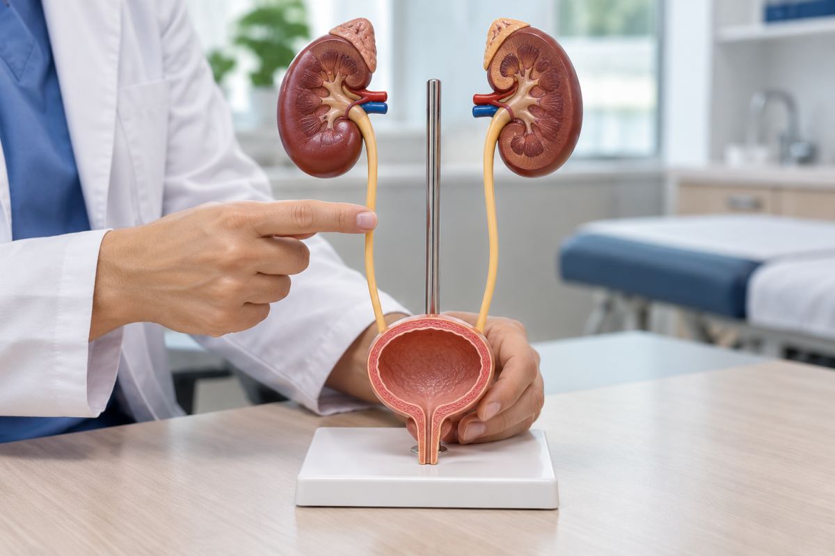
The ureters are the tubes that carry urine from the kidneys to the bladder. They are distinct from the urethra, which carries urine out of the body. When drainage is impaired, the renal pelvis—the collecting area inside the kidney—can become enlarged. This dilation is called hydronephrosis and can affect one or both kidneys. It is a finding that calls for investigation of the underlying cause; it does not, on its own, establish a particular congenital abnormality.
One recognized condition is ureteropelvic junction obstruction, a blockage where the renal pelvis joins the ureter. According to the American Academy of Pediatrics, in babies it is usually associated with narrowing at that junction. In older children and adolescents, a blood vessel crossing the ureter may also interfere with urine flow.
The condition may be detected on a prenatal ultrasound or investigated after a urinary tract infection. Symptoms can include abdominal pain, discomfort in the side or back, nausea and vomiting. How severe the symptoms feel is no substitute for assessing how well the kidney is working.
Another finding is megaureter, meaning an enlarged ureter. The University of California, San Francisco emphasizes that this, too, is a description rather than a complete diagnosis. It may result from a blockage near the bladder or from urine flowing backward into the ureter, known as reflux. It can also occur without either problem. In some cases, obstruction and reflux occur together.
These distinctions affect treatment. An enlarged ureter does not always require surgery: some childhood cases improve as the child grows. Others require an operation if infections recur or kidney function declines. Grouping them under a phrase such as “misshapen ureter” can therefore obscure crucial differences.
There is also an uncommon abnormality called a retrocaval ureter, in which part of the tube passes behind the inferior vena cava, a major vein. This route can interfere with drainage. The news reports reviewed do not provide evidence that this was Daikichi’s condition.
In 2024, a team at Isahaya General Hospital in Nagasaki reported a robot-assisted reconstruction of a retrocaval ureter in IJU Case Reports. The operation involved dividing the ureter, moving it in front of the vein and joining it back together. The case illustrates one treatment option, but the authors acknowledged important limitations: it involved only one patient and a short follow-up period. It cannot establish that everyone with the condition will have the same outcome.
The causes therefore depend on the specific abnormality. Ureteropelvic junction obstruction can involve developmental factors within the junction itself as well as external factors. There is no single cause that explains every abnormality described informally as a curve or a twist.
Nor is there one test that can settle every case. European Association of Urology guidelines recommend considering the clinical course alongside imaging and kidney function studies. Ultrasound shows dilation; a diuretic renogram helps assess drainage and each kidney’s share of overall function. Other tests may be used in particular circumstances. Decisions depend on the findings taken together.
The consequences can range from temporary dilation to impaired kidney function. The US National Institute of Diabetes and Digestive and Kidney Diseases, or NIDDK, warns that hydronephrosis in infants can be associated with urinary tract infections, stones and chronic kidney disease. Many mild cases, however, improve on their own.
To put the finding in perspective, the institute states that fetal hydronephrosis is diagnosed in one or two out of every 100 pregnancies and that roughly half of those cases resolve before birth. That figure covers prenatal hydronephrosis from different causes. It is not a worldwide estimate of how common a “curved ureter” is.
Estimates for ureteropelvic junction obstruction also differ: the American Academy of Pediatrics cites about one in 1,000 children, while the European guidelines give a figure of one in 1,500. These should be presented as estimates for that particular condition, not as a global count of all congenital ureter abnormalities.
When a narrowed ureteropelvic junction needs surgery, a common procedure is pyeloplasty: the affected segment is removed and the connection reconstructed to allow urine to drain more freely. Osaka University explains that the aim is to protect kidney function and prevent infections, pain or blood in the urine.
The operation can be performed through different approaches, including laparoscopic and robot-assisted surgery. The choice depends on the patient’s anatomy, body size and clinical circumstances. An internal ureteral stent may be placed temporarily between the kidney and bladder to help urine drain during recovery.
There is no mortality rate that can meaningfully be assigned to the term “curved ureter.” The risk depends on the obstruction, kidney damage and complications. In severe cases, a kidney infection can cause scarring, kidney failure or sepsis. Sepsis is an extreme response to infection that can lead to organ failure and death without prompt treatment, according to MedlinePlus, a service of the US National Library of Medicine.
The combination of obstruction and infection warrants particular attention. European guidelines on urinary stones classify an obstructed kidney with signs of infection, or a lack of urine production, as a urological emergency. Treatment may require antibiotics and urgent drainage through a ureteral stent or a nephrostomy tube, which drains urine from the kidney.
Fever accompanied by pain in the side, particularly in someone with a known obstruction, requires prompt assessment. Investigating these abnormalities and maintaining follow-up are intended to identify when an anatomical difference is impairing drainage, so that treatment can begin before the damage progresses.